New York American College of Emergency Physicians

Geoff W. Jara-Almonte, MD

Geoff W. Jara-Almonte, MD

Associate Residency Director, Department of Emergency Medicine Icahn School of Medicine at Mount Sinai

Bronchiolitis

With two hours to go on a busy evening shift, a new patient pops up on the board. The line on the EMR reads 9 mo M. CC: Breathing Problem VS: T 37.9 RR 38 HR 148 SpO2 95%. Everyone else is busy, so you decide to pick up the kiddo.

The parents tell you their child was born full term and had no complications at birth; his vaccines are up to date. Parents noticed that two days ago he started with rhinorrhea and nasal congestion and then one day ago began to cough. They feel like his cough has just been worsening and thought he was breathing hard tonight so elected to bring him in. On exam ou find a well-developed baby sitting upright in the parent’s lap. He is interactive and curious. On lung exam you note bilateral wheezing and scattered crackles. As you finish your exam you start to come to a working diagnosis.

Bronchiolitis is a clinical diagnosis that, according to the American Academy of Pediatrics, is recognized as “a constellation of signs and symptoms occurring in children younger than two years, including a viral upper respiratory tract prodrome followed by increased respiratory effort and wheezing.”1

We usually consider the diagnosis in young children presenting with acute respiratory infections who have both upper and lower respiratory tract involvement and present in the typical “RSV season”. There is a broad differential to consider including bacterial and viral pneumonias, influenza, reactive airway disease and croup. Other less common conditions to consider include airway foreign bodies and congestive heart failure.

Differentiating bronchiolitis from reactive airway disease in the setting of a concomitant URI is probably the most challenging discrimination to make. Repeated prior episodes of wheezing, formal diagnosis of asthma, personal history of atopy, or a strong family history of asthma or atopy may make one more suspicious for reactive airway disease. Bacterial pneumonia classically presents with a more abrupt onset of high fever and lower respiratory tract signs and symptoms whereas in bronchiolitis fever is typically lower grace (< 38.3) and lower respiratory tract findings occur only after a URI prodrome. Influenza too typically has a higher grade fever and more systemic involvement; lower respiratory tract findings are uncommon however. Croup may also present with URI that progresses to increased respiratory effort and tachypnea. In the case of croup, however, respiratory distress is due to upper airway obstruction rather than lower. Differentiation can be made based on the presence of stridor and characteristic “brassy” cough in croup as opposed to wheezing in bronchiolitis.

An aspirated airway foreign body is a rare cause of wheezing. There may be a history of choking, but not always. Typically wheezing is focal. Onset is classically abrupt and fever is absent in acute aspiration. Pulmonary edema due to congestive heart failure may present as wheezing. A history of progressively poor feeding, diaphoresis with feeding, and poor weight gain may increase suspicion. On exam one may note hepatomegaly or heart murmur.

On further history the parents deny any family or personal history of atopy. They confirm that there has been no highgrade fever and confirm a progression of symptoms typical of bronchiolitis. Prior to the development of URI symptoms a few days ago he had been in his usual state of health with no concerns. The parents also report that at daycare several children have been ill with “a chest cold”

At this point you feel fairly confident with the diagnosis of bronchiolitis but wonder if you should perform additional testing.

Bronchiolitis is a clinical diagnosis and generally does not require ancillary studies to confirm the diagnosis or exclude complications in patients who are not critically ill. Children will commonly have abnormalities on chest x-ray including peribronchial cuffing, atelectasis, or hyperinflation, however their presence has not been shown to correlate with progression to severe disease. X-ray should probably be reserved for cases in which there are clinical features that support an alternative diagnosis.

Viral testing is commonly performed, though strong evidence to support its patient-level utility is lacking. Respiratory Syncytial Virus (RSV) is the most common causative etiology of bronchiolitis, accounting for about 75% of cases. A multitude of other viruses including rhinovirus, metapneumovirus, adenovirus, non-sabre coronaviruses and parainfluenza virus have been implicated as well. However, in most emergency department (ED) settings only RSV testing (sometimes in combination with influenza and SARS-CoV2) is readily available. A negative RSV test does not exclude bronchiolitis.

Some evidence does suggest that RSV, as compared to rhinovirus, has a typical course of illness and so confirmation of infection may allow for more accurate patient counseling. In addition, many inpatient services will “cohort” RSV positive patients as an infection control measure. Confirmation of RSV infection may reduce antibiotic use as well. For these reasons testing is commonly performed in the ED.

In some centers a respiratory virus panel that tests for a multitude of other viral pathogens may also be obtained. In general identification of a specific viral pathogen does little to alter management. In addition, prolonged viral shedding after resolution of an acute infection may result in a positive result that is unrelated to the acute presentation, thus complicating the diagnostic process. Additional discussion of the potential benefits and pitfalls of respiratory viral testing are beyond the scope of this discussion.

In general, additional studies such as blood count and serum chemistries are not helpful except in the setting of critical illness.

You decide to obtain an RSV test, which returns positive. You discuss with the parents that their child has classic RSV bronchiolitis and discuss the typical clinical course. The parents tell you that a friend’s child was recently admitted to the hospital for bronchiolitis and wonder if that is necessary for their son.

The clinical severity of bronchiolitis is highly variable. In the vast majority of cases it is a relatively benign selflimited disease. However severe disease may occur and can be complicated by respiratory failure, apnea or dehydration. Disposition decisions should be made based upon the current clinical severity, anticipated clinical course, host factors that affect potential for decompensation, availability of follow-up and caregiver comfort and resources.

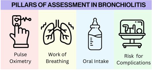
A variety of clinical scoring systems have been developed that assess severity of illness and risk of progression, however none has demonstrated definite superiority. When formulating a disposition plan the emergency clinician should consider four essential questions:

1) Is there hypoxia?

2) Is there significantly increased work of breathing or respiratory distress?

3) Is the patient able to maintain adequate oral intake?

4) Is the patient at high risk for complications or rapid / severe progression of disease?

Inpatient management is generally indicated for children with hypoxia. The exact level at which admission is indicated is not clear. Many providers would be comfortable with outpatient management if the SpO2 > 95%, and would admit if SpO2 < 91. Between 90% and 95% there exists a degree of variability in practice. The AAP guidelines allow for managing those children with SpO2 >90 without supplemental oxygen as a treatment option; whether they should be routinely admitted for monitoring of the oxygen saturation is not clear. Some authors have suggested that infants with isolated hypoxia who are not in respiratory distress may be managed with home oxygen.

Work of breathing is a clinical assessment that requires a careful and attentive physical exam. A large body of literature has documented the inaccuracy of triage respiratory rate; it is imperative that the emergency clinician assess this themself. Some studies have identified tachypnea as a risk factor for progression to respiratory failure. Examination for accessory muscle use must be performed with the child undressed; it is impossible to assess for subtle subcostal or intercostal retractions while a onesie is on. Careful attention should be paid to detect tracheal tugging, grunting, nasal flaring and head bobbing; presence of these signs may portend progression toward respiratory failure.

A reasonably careful feeding history should be obtained: how much has the patient fed in the last 12 – 24 hours, how much would they usually have fed and what is the adequacy of urine output as measured by number and fullness of wet diapers. It is not unusual for children to feed less while ill. Older children who are taking solids may have decreased or ceased solid intake. This may be distressing to parents but is generally well tolerated so long as there is adequate fluid intake. In younger infants, fluid intake may fall off and parents may report providing smaller more frequent feedings. Again, this is generally sufficient. It may be reasonable to observe one or two feedings in the ED to get a better sense of the adequacy of oral intake. Those patients who manifest clinical signs of dehydration or are observed to be unable to tolerate sufficient feeds may require admission for IV or enteral hydration.

The most serious complications of bronchiolitis are respiratory failure and apnea. Several host factors have been described that may be associated with development of either complication. These include prematurity, young age, bronchopulmonary dysplasia, hemodynamically significant congenital heart disease, immunocompromised state. In general, a lower threshold should be had for admission in these special populations. Special attention must be paid to age. Full term children less than eight weeks life or preterm children less than 48 weeks post-conceptual age seem to be at increased risk for apnea; many emergency clinicians will admit very young children for monitoring even in the absence of other indications.

When arriving at a disposition decision it is imperative to consider the current stage of illness; RSV bronchiolitis tends to follow a predictable clinical course with severity of respiratory symptoms peaking on day 3 – 5. Rhinovirus bronchiolitis seems to have a shorter duration. Wheezing may persist for over a week, and full resolution may take up to 21 – 28 days. Understanding of this clinical course is important for formulating a disposition plan. For example, a child who is borderline in terms of severity, but on day five of illness and can see their pediatrician in 12 hours may be a more suitable candidate for outpatient management than a child who presents with the same severity of disease on day three of illness leading into a three-day weekend.

A careful feeding history reveals the child has been taking smaller bottles, but is doing so more frequently. He feeds once in the ED taking about six oz of formula. The parents feel his urine output is at baseline. He has a pediatrician he can see in the morning and after a long discussion with parents you determine he can be managed as an outpatient. The parents tell you that another friend of theirs has a child with asthma who frequently wheezes and has to get breathing treatments. They wonder if they need to do the same thing since their child is breathing.

Asthma and bronchiolitis are characterized by decreased diameter of the small airways, airflow obstruction and wheezing, though the mechanisms are different. In asthma reversible constriction of the bronchiolar smooth muscle plays a predominant role. In bronchiolitis the decrease in bronchiolar cross-sectional area is due to edema from infection and collection of cellular debris caused by bronchiolar endothelial sloughing; smooth muscle constriction does not play a large role in the pathogenesis of airflow obstruction.

Studies examining the role of bronchodilators have failed to demonstrate benefits other than transient improvement in severity scores. The AAP strongly recommends against use of bronchodilators or steroids in bronchiolitis. It is important to remember these guidelines do not apply to children with recurrent episodes of wheezing, asthma, or pre-existing pulmonary pathology. An approach to children with a history of recurrent wheezing or a high suspicion for reactive airway disease may be individualized based on clinical circumstances and history.

The role of bronchodilators in steroids in recurrent episodes of bronchiolitis is less clear, as clinical signs and symptoms may overlap. It may be worthwhile to consider additional historical factors, such as family, history of asthma, atopy and occurrence of wheezing episodes outside the context of clinical bronchiolitis. If these factors are present, they may prompt a trial of bronchodilator therapy. The utility of steroids for viral associated wheezing is unclear.

One reasonable therapy to provide in the ED is nasal suctioning. Upper airway secretions can contribute to respiratory distress and lead to difficulty with feeding. A trial of nasal suctioning and observation for improvements in work of breathing and feeding may confer significant benefit, especially in younger patients. If successful, patients can be educated and provided with a bulb suction syringe. Additional nasal aspiration devices can also be obtained at a pharmacy.

Absent a high suspicion for secondary or concomitant bacterial infection, antibiotics are not indicated.

After discharging the patient and his family, the patient’s nurse comes back to you to ask about what you would have had to do if the child had been more severely ill. “If there’s nothing we can really do to treat these kids, what do you do when they’re really sick?” he asks.

Hypoxemia in children with bronchiolitis can generally be managed with supplemental oxygen delivered by nasal cannula, blow-by or tent. In the case of severely increased work of breathing non-invasive ventilatory modalities may be beneficial. Continuous positive airway pressure, bilevel positive airway pressure and heated humidified high-flow nasal cannula have all been studied. While high-quality prospective evidence and definitive guideline recommendations are lacking, most emergency clinicians will attempt non-invasive ventilation for infants with bronchiolitis complicated by hypoxemia and respiratory distress who have adequate mental status and no complications. In general, high-flow is employed as first-line therapy given its ease of use and the fact it is generally well-tolerated.

While well-appearing children can be managed without significant use of ancillary studies, those who are critically ill probably warrant additional studies including blood count and serum chemistries. Blood gasses are commonly checked as well. It is reasonable to obtain a chest x-ray to evaluate for complications such as pneumothorax or evidence of bacterial pneumonia.

Your patient is discharged home and follows-up with the pediatrician the next day. When you call to follow up after a few days the parents report the child is doing well and is back to normal except for a mild cough. They thank you for your help and are happy to have had your care!

Wrap Up and Key Points

  • Bronchiolitis is a clinical syndrome recognized by signs and symptoms of lower respiratory tract involvement that develops after a characteristic prodrome of upper respiratory symptoms in a child less than two years of age.
  • In a typical presentation the diagnosis can be made clinically without routinely obtaining ancillary studies.
  • The differential is broad and care should be made to ensure there is not another cause of the patient’s presentation that would demand specific therapy.
  • Care is primarily supportive. There are no disease-specific treatments. Therapies aimed at reducing airflow obstruction are generally not indicated.
  • Careful attention should be paid to assess for complications including hypoxemia, respiratory insufficiency / failure and dehydration.
  • Assessment for host factors that may increase risk of progression to more severe disease should be made.
  • Disposition decisions should take into account the clinical stage of illness and anticipated clinical course
  • A trial of non-invasive ventilatory therapy is reasonable for many patients with respiratory insufficiency or failure.

References

1. Ralston, Shawn L., et al. “Clinical practice guideline: the diagnosis, management, and prevention of bronchiolitis.” Pediatrics 134.5 (2014): e1474-e1502.