New York American College of Emergency Physicians

Penelope C. Lema, MD RDMS FACEP

Penelope C. Lema, MD RDMS FACEP

Vice Chair, Faculty Affairs Associate Professor, Department of Emergency Medicine Columbia University Vagelos College of Physicians & Surgeons

Guest Author Tyler Wen, MD

Guest Author Tyler Wen, MD

PGY-3 Emergency Medicine Resident NYP Columbia Cornell Emergency Medicine Residency

Guest Author Miles Gordon, MD FPD-AEMUS

Guest Author Miles Gordon, MD FPD-AEMUS

Assistant Professor of Emergency Medicine Department of Emergency Medicine Columbia University Vagelos College of Physicians and Surgeons

Lend me Your Ear: Diagnosis of a Preauricular Abscess

Case Presentation

A 50-year-old female with a past medical history of a right preauricular sinus tract, previously complicated by abscess requiring aspiration presented to a community Emergency Department (ED) with four days of worsening right ear pain associated with chills. She had initially been evaluated on day one of symptoms at an urgent care and was prescribed ciprofloxacin without improvement. This was followed by the addition of cephalexin at a second visit, however her pain significantly worsened and therefore presented to the ED for a third visit.

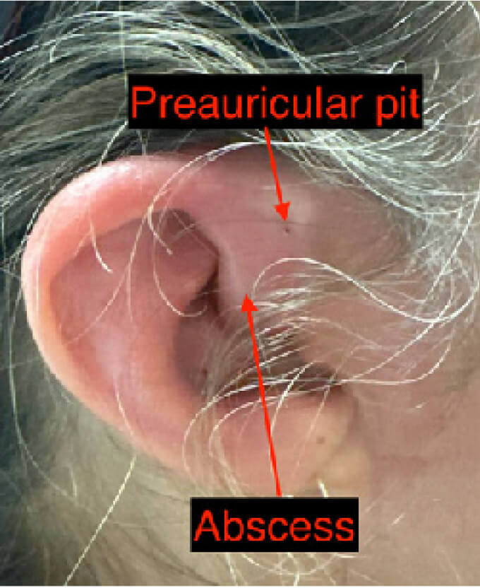
On exam, there was moderate swelling, erythema and warmth of the superior aspect of the right helix and crus of the helix, with induration superior to the tragus (Figure 1). Examination for fluctuance was limited due to significant hyperesthesia of the area. A point-of-care ultrasound was performed with a high-frequency linear probe with a copious amount of gel to prevent any additional pressure to the area. The ultrasound demonstrated a 1.8cm x 0.8cm x 0.7cm anechoic collection just superior to the tragus (Figure 2). We identified an optimal point of needle entry after visualizing surrounding vasculature on color doppler (Figure 3). An 18g needle was used to perform an aspiration and drainage, which removed 1cc of purulent material with significant improvement in pain. The patient was treated with vancomycin and piperacillin/ tazobactam and was admitted for IV antibiotics and observation. Ultimately, a wound culture resulted as Enterococcus faecalis infection and the patient was successfully discharged with oral antibiotics.

Discussions

Preauricular sinuses are uncommon congenital malformations that can manifest as superficial dents, dimples or openings typically anterior to the crus of the helix.1 Preauricular sinuses usually remain asymptomatic for most patients; however, these sinuses are at risk of developing inflammation and infection, with typical early symptoms including pain, erythema, warmth and edema of the preauricular region.2 If left untreated, underlying infections can develop into abscesses that can affect nearby structures such as the facial nerve or the auricular cartilage itself, potentially leading to facial nerve palsy and perichondritis, respectively.3 The relative rarity of preauricular sinus abscesses in the population at large can also lead to underdiagnosis of the condition, as it could be clinically mistaken for otitis externa, perichondritis or superficial cellulitis. As a result, prompt and accurate identification of preauricular sinus infections are key to preventing morbidity.

The majority of preauricular sinus infections are caused by Staphylococcal species, with other less common culprit organisms belonging to the Streptococcus, Proteus, and Peptococcus families 2. Antibiotics with coverage of these species, such as cephalexin or amoxicillin-clavulanate are appropriate first-line medications. If an abscess is identified, needle aspiration and drainage or incision and drainage are indicated. In this case, an Enterococcal species was isolated as the causative organism, possibly through fecal-facial contamination.

Ultrasonography of preauricular sinus abscesses can reveal pockets of purulence that may then be targeted by needle aspiration. In our case, the patient’s preauricular abscess appeared as a pocket of heterogeneous echogenicity just below the skin, allowing drainage without needing to violate nearby cartilage. By utilizing POCUS, we were able to adequately characterize the underlying pathology that led the patient to seek a medical evaluation three times in one week while avoiding CT irradiation. This case therefore illustrates the utility of bedside POCUS to identify preauricular sinus infections and guide subsequent management.

Indications

  • Erythema
  • Fluctuance
  • Preauricular pain
  • Preauricular pit
  • Swelling
  • Warmth

Pitfalls and Limitations

  • Pain in affected areas may limit the ability to perform bedside POCUS; in these cases it may be useful to use copious amounts of gel to minimize pressure-induced discomfort or to treat with a topical anesthetic prior to imaging
  • Patients with prominent preauricular lymph nodes, aberrant vasculature or a parotid mass could appear similar to anechoic abscesses on POCUS. Using color flow or pulsed wave Doppler modes can help clarify the identity of the visualized structures.
Figure 1: Image of outer ear with preauricular pit and adjacent area of fluctuance.
Figure 2: Ultrasound of a preauricular sinus abscess in longitudinal view, with internal heterogeneous echogenicity using a linear transducer.
Figure 3: Color flow Doppler illustrating optimal area of needle entry (dashed arrow).

References

  • Matev B, Lyutfi E, Stoyanov GS, Sapundzhiev NR. Preauricular Sinus: A Tale of Forgetful Rediscovery. Cureus. 2020 Jun 28;12(6):e8885. doi: 10.7759/cureus.8885. PMID: 32742852; PMCID: PMC7388807.
  • Scheinfeld NS, Silverberg NB, Weinberg JM, Nozad V. The preauricular sinus: a review of its clinical presentation, treatment, and associations. Pediatr Dermatol. 2004 May-Jun;21(3):191-6. doi: 10.1111/j.0736-8046.2004.21301.x. PMID: 15165194
  • Adegbiji WA, Alabi BS, Olajuyin OA, Nwawolo CC. Presentation of preauricular sinus and preauricular sinus abscess in southwest Nigeria. Int J Biomed Sci. 2013 Dec;9(4):260-3. PMID: 24711764; PMCID: PMC3884798.