New York American College of Emergency Physicians

Geoff Jara-Almonte, MD

Geoff Jara-Almonte, MD

Associate Residency Director Department of Emergency Medicine Icahn School of Medicine at Mount Sinai NYC H+H Elmhurst Hospital Center

Alexandra Bourlas, DO

Alexandra Bourlas, DO

Clinical Instructor in Emergency Medicine Weill Cornell Medicine Attending Physician NewYork-Presbyterian

A Neonatal Resuscitation Refresher

It’s likely every emergency medicine (EM) physician’s worst nightmare (or at least one of the many): An imminent or precipitous delivery. Two patients for the price of one and so much can go wrong. But as is an ever-running theme in our specialty, we must be prepared for anyand everything that comes through our doors.

The good news is if this does occur on shift, the likelihood you will need to do much more for this new addition to the world apart from a quick neonatal evaluation and cord clamping is low. Studies suggest only about 10% of all newly born infants require some breathing assistance at birth, with extensive resuscitation necessary in up to 1%. Even with this relative rarity, neonatal mortality in the US is about 4 per 1000 live births and many EM physicians feel they need more exposure to neonatal resuscitation. So, without further ado, a refresher on the very important topic of neonatal resuscitation. (We’ll leave the Moms for another day).

Understanding Burnout

Burnout is a complex and multifaceted phenomenon characterized by emotional exhaustion, depersonalization and a reduced sense of personal accomplishment.2,3 The fast-paced, high-stakes environment of emergency medicine has been associated with near and long term effects on providers including the development of burnout among physicians.1 The constant pressure, long hours and exposure to traumatic events can take a toll on their mental and emotional well-being, with past work finding elevated risk for the development of psychological and even physiological changes such as blood pressure, poor sleep, and anxiety.5

Similarly, clinician researchers, who navigate the delicate balance between clinical practice and research endeavors, face unique challenges. The pressures to publish, secure grants and juggle clinical responsibilities can lead to high degrees stress and burnout. The parallel demands of clinical and research roles make it imperative to recognize burnout as a shared concern in both spheres of such clinician-investigators.

Preparation

In the rare instance you receive a pre-hospital notification for a neonatal resuscitation, the first step is preparation. Turns out, a lot of equipment and resources may be necessary to support this patient (see below). Most importantly, however, make sure to have a neonatal radiant warmer turned on and warmed up and ensure you have a compressed air source, oxygen blender with flow meter, neonatal pulse oximeter, bag-valve mask and laryngeal mask available in your resuscitation bay. Familiarize yourself with this equipment early and often. The last thing you want at that critical moment is to have to figure out how to operate any of it. And though all resuscitative gear has some level of importance, the greatest resource any of us can have is the help of our respiratory therapy and NICU comrades.

Once the patient(s) arrives, quickly perform an assessment of the maternal and fetal history. What is the gravidity and parity? When was her last menstrual period or does she know her estimated gestational age (EGA)? What’s the patient’s prenatal history? Did they have gestational diabetes or hypertension in pregnancy? Are there any concerns for infectious serologies or known genetic anomalies? Did the patient have any prenatal care? Is she experiencing a fever or have there been prolonged rupture of membranes? Was there meconium-stained amniotic fluid? If a precipitous delivery has occurred, there may not be time to ask all of this up front. Perhaps the mother doesn’t know how pregnant she is, or that she was even pregnant at all. In cases like these, move onto the fetal assessment and ask questions along the way. The fetal physical exam can provide clues to help you approximate an EGA if the mother is unsure. For example, if eyes are spontaneously opening, the neonate is likely term or >37 weeks EGA. If the eyes are fused shut, they’re more likely between 20-28 weeks. Other signs of prematurity, include thin, translucent and sticky skin and shiny, smooth soles of the feet without creasing. Lanugo, the fine hair covering a fetus acquires in utero, appears around 20-25 weeks, is abundant by 28 weeks and normally is shed at about 33-36 weeks. All of this information will be helpful in guiding your resuscitative efforts, including the unfortunate decision of when to terminate those efforts.

Resuscitation

Is the infant breathing/crying spontaneously with good tone? If so, get that baby to mom and complete your assessment while they’re enjoying skin-to-skin bonding. Make sure to provide warmth via skin-to-skin or blankets, clear the nose and mouth with bulb suction if there are signs of obstructed breathing, dry the baby and provide ongoing assessment of respiratory effort and tone. In cases like these, you can allow for delayed cord clamping of 1-3 minutes in both term and healthy pre-term neonates, as it can reduce the need for blood transfusion, increase neonatal iron stores and may improve survival. For those newborns requiring immediate resuscitative intervention, the cord should be clamped and cut and the baby should be moved to the isolette for further evaluation and care.

The First 30 Seconds – The T- ABCs

The goal is to complete an initial clinical assessment and provide quick and easy interventions in the first 30 seconds or less after birth, starting with the T-ABCs: Temperature, Airway, Breathing, and then Circulation. Newborns showing poor tone or poor respiratory effort should be placed under a preheated radiant heat source (e.g. radiant warmer) and gently dried and stimulated with a warm towel. If you have a very-low birth-weight newborn or one <29 weeks EGA, they should be placed in a polyethylene bag. Plastic food wrap or a food-grade 1-gallon plastic bag may also be used. Take care to avoid hyperthermia, which may precipitate apnea and worsen hypoxic-ischemic injury in depressed infants.

If the infant is not breathing, dry and provide stimulation by rubbing the back or tapping their foot a few times. If there is still no response, open the airway using a jaw thrust and place them in the sniffing position with towels beneath the shoulders. If there appears to be obstruction from amniotic fluid, gently suction the nose and throat with a bulb or 8F catheter. Current guidelines advise against routine deep suctioning of the newborn as tracheal suctioning can cause reflex bradycardia and apnea. If none of these efforts are successful, time to move on to the next 30 seconds of interventions.

The Next 30 Seconds – Heart Rate and Positive Pressure Ventilation

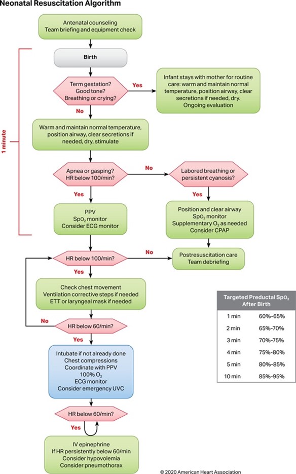
If the neonatal heart rate is >100 bpm, but they are persistently cyanotic or with labored breathing, open the airway and suction nose and mouth. Attach a pulse oximeter to the right hand or wrist to measure a preductal SpO2 and apply supplemental oxygen to achieve targeted preductal O2 saturation goals (see below). If the heart rate is <100 bpm, or if the neonate is gasping or remains apneic after the initial steps above, it’s time to start positive pressure ventilation (PPV).

PPV can be administered via a few different types of devices, including self-inflating bags, flow-inflating bags and T-piece resuscitators. Check with your hospital’s central supply or NICU to determine what is available to you and familiarize yourself with its operation. Flow-inflating bags are typically preferred by neonatal resuscitationists, but they take more training and experience to properly operate. In inexperienced hands, self-inflating bags are superior. Start PPV with a ventilation rate of 40-60 breaths/min. Care must be taken to avoid volutrauma and barotrauma. Ensure that a neonatal ( not child- or adult-sized) Bag Valve Mask (BVM) is used. Provide just enough volume to achieve visible chest rise. Infant BVMs include a manometer and pop-off valve that typically limits peak inspiratory pressure (PIP) to 20 mmHg when active. Attention must be paid to ensure excessive PIP is not provided. Typically, a PIP of 20 cmH2O is usually sufficient, but as high as 30 – 40 cmH2O may be required for the first breath or two to overcome the surface tension in fluid-filled alveoli. Exposure to excessive inspiratory pressures can cause pneumothorax and compromise your resuscitation. Additionally, start resuscitative efforts on room air as excessive oxygenation is associated with increased mortality. As in most resuscitations with children, neonatal resuscitation.

As in most resuscitations with children, neonatal resuscitation requires attention to airway and breathing before circulation. Bradycardia (even when extreme) is usually a result of respiratory failure. Before compressions or resuscitative medications, first make sure you are effectively ventilating your patient. Delaying PPV can increase patient mortality and prolongs hospitalization. Watch for chest rise and an increase in heart rate, which is the most sensitive indicator for successful ventilation – An increase in heart rate within 5-10 breaths suggests you are effectively ventilating your patient. Utilize your 3-lead EKG to gauge your efforts.

Permissive Hypoxia

You’re seeing a rise in heart rate, but the neonate’s pulse oximeter is only reading 65%. Don’t panic! Fetal circulation undergoes many physiologic changes at birth. At this point, the baby is relying on its own lungs to supply oxygenated blood for systemic circulation. The gradual increase in oxygen saturation (SpO2) within the first few minutes of life reflects this transition to extrauterine life . This transitional process normally occurs in a few minutes and, rarely, can take up to hours or even days. By the first minute of life, the normal neonate’s SpO2 is 60% to 65%, increasing steadily by 10% about every 2 minutes. Only after roughly 10 minutes of life will the full-term, healthy neonate exhibit an SpO2 of >85%.

If you aren’t seeing a response to PPV, attempt the following corrective steps prior to moving to more advanced resuscitative efforts. This is commonly known by the mnemonic: MR. SOPA

M – Mask (adjust to improve seal)
R – Reposition head to open airway
S – Suction (the mouth first, then the nose)
O – Open the mouth (jaw thrust)
P – Pressure (increase it until chest rise is noted – Max PIP 40cmH2O)
A – Airway control (aka intubate!)

60 Seconds and Beyond – Advanced Resuscitation

Yes, you understood that correctly. Our goal is to attempt all of the above within the first 60 seconds of life. If the neonatal heart rate is <60 bpm despite your best efforts, it’s time to start chest compressions. This should be done at a rate of 3 compressions to 1 breath for a total of 30 breaths and 90 compressions (120 events) per minute. The two-thumb technique seems to be superior in generating greater peak systolic pressures. However, the two-finger technique may be more practical if a colleague is simultaneously attempting umbilical vessel catheterization. At this time, you’ve likely intubated the patient and are ventilating them with an FiO2 of 100%. Once the neonatal heart rate has exceeded 60 beats/min, you can stop chest compressions and focus on ventilation, increasing the ventilation rate to 40-60 breaths/min. Slowly wean PPV when the heart rate exceeds 100 bpm and the newborn has begun to breathe spontaneously.

If the heart rate has not improved despite adequate ventilation and compressions for 45-60 seconds, it’s time for vascular access and epinephrine. IV/IO access can be obtained, but consider umbilical vein catheterization if access is difficult to obtain or if you think blood transfusion or prolonged infusions may be necessary. As is a common theme with this topic, familiarizing yourself with an umbilical vein catheterization is paramount for success. Epinephrine can be given 0.01 to 0.03 milligram/kg IV/IO or endotracheal dosing 0.05 to 0.1 milligram/kg. Volume expansion should also be considered when there is known or suspected blood loss (e.g. pallor, poor perfusion or weak pulses). Administer 10 mL/kg of 0.9% saline normal saline or O-negative blood – slowly over 3-5 minutes. The slow infusion is especially important in premature infants who are at risk for intraventricular hemorrhage. You may also want to check for hypoglycemia and pneumothorax at this time. Hypoglycemia is associated with adverse outcomes following birth asphyxia. Check for hypoglycemia (<50 mg/dL) and correct as needed with 5mL/kg IV push of D10 or a D10 infusion at 4-7mg/kg/min. You can assess for pneumothorax quickly utilizing the transillumination test – place a light source on the chest wall. A translucent chest wall and visible light source is suggestive of pneumothorax. If there is high clinical suspicion for pneumothorax, you may also perform needle aspiration with a saline-filled syringe in the 2nd or 3rd intercostal space at the mid-clavicular line. When air bubbles are present in the syringe, you have entered the pneumothorax and can evacuate it.

Up to 90% of newborns will require only basic maneuvers such as warming, drying and stimulation. And when a newborn requires resuscitation, there may be abnormalities in major organ systems at play. The list of potential organ system dysfunctions and considerations for post-resuscitative care is lengthy and out of our scope of practice. Hopefully by this time in your efforts, the NICU team has taken over and/or you have initiated transfer to a tertiary or quaternary care center.

Termination of Efforts

In all true medical emergencies, advanced preparation can mean the difference between good and bad outcomes – life or death. But sometimes we are unsuccessful despite our best efforts, and our patients die. Just as it is important to know when and how to do everything you can to save a life, it is equally as important to know when to stop. EGA, signs of life during resuscitative efforts, length of active resuscitation and presence of severe congenital anomalies all must be considered when making this difficult decision. Peri-viability is typically noted between 22-24 weeks EGA and data suggests that a neonate with no signs of life and an EGA of <22 weeks has a 0% chance of survival, with comfort care rather than resuscitation, the recommended course of action. Additionally, newborns with no signs of life after 10 minutes of continuous resuscitation are virtually certain to suffer severe morbidity and/or mortality if vital signs are restored. If there are no signs of life despite performing all steps of resuscitation approximately 15-20 minutes after birth, we have an ethical responsibility to begin discussing a change in goals of care and cessation of our efforts with the team and family.

Neonatal resuscitation is a rare and highly stressful event. The last thing any emergency medicine physician wants is to be unprepared when the stakes are this high. Luckily, the vast majority of deliveries are uncomplicated and require very little from us. In the rare event where more intervention is necessary, remember that anticipation, preparation and effective team communication are the keys to success. Focus on providing high quality respiratory support, considering airway and breathing before circulation, using the neonate’s heart rate as your guide. And get help early from whomever is willing and able.

The American Heart Association (AHA) and Neonatal Resuscitation Program (NRP) have algorithms to guide clinicians through the process step-by-step.

References

1. Collin MF. Resuscitation of Neonates. In: Tintinalli JE, Ma O, Yealy DM, Meckler GD, Stapczynski J, Cline DM, Thomas SH. eds. Tintinalli’s Emergency Medicine: A Comprehensive Study Guide, 9e. McGraw Hill; 2020. Accessed March 23, 2023. https://accessmedicine-mhmedical-com. clinicalproxy.libr.ccny.cuny.edu/content.aspx?bookid=2353§ionid= 219643848

2. Aziz K, Lee HC, Escobedo MB, et al. Part 5: Neonatal Resuscitation: 2020 American Heart Association Guidelines for Cardiopulmonary Resuscitation and Emergency Cardiovascular Care. Circulation. 2020;142(16_suppl_2):S524-S550. doi:10.1161/CIR.0000000000000902

3. Vo AT, Cho CS. Neonatal Resuscitation in the ED. Pediatric Emergency Medicine Practice. December 1, 2020. Accessed April 3, 2023. https:// www.ebmedicine.net/topics/critical-care/neonatal-resuscitation

4. Ersdal HL, Mduma E, Svensen E, Perlman JM. Early initiation of basic resuscitation interventions including face mask ventilation may reduce birth asphyxia related mortality in low-income countries: a prospective descriptive observational study. Resuscitation. 2012;83:869–873. doi: 10.1016/j.resuscitation.2011.12.011

5. Berazategui JP, Aguilar A, Escobedo M, Dannaway D, Guinsburg R, de Almeida MF, Saker F, Fernández A, Albornoz G, Valera M, Amado D, Puig G, Althabe F, Szyld E; ANR study group. Risk factors for advanced resuscitation in term and near-term infants: a case-control study. Arch Dis Child Fetal Neonatal Ed. 2017;102:F44–F50. doi: 10.1136/ archdischild-2015-309525