New York American College of Emergency Physicians

Penelope C. Lema, MD RDMS FACEP

Penelope C. Lema, MD RDMS FACEP

Vice Chair, Faculty Affairs Associate Professor, Department of Emergency Medicine Columbia University Vagelos College of Physicians & Surgeons

Guest Author Miles Gordon, MD

Guest Author Miles Gordon, MD

Assistant Professor of Emergency Medicine Columbia University Department of Emergency Medicine Vagelos College of Physicians and Surgeons

Guest Author Tyler Wise, MD

Guest Author Tyler Wise, MD

PGY-3, Emergency Medicine Resident New York – Presbyterian Columbia Cornell Emergency Medicine Residency

Abdominal Pain With a “Negative” CT…Sonographic ‘Tic Tips

Case

A 56-year-old female with a significant past medical history of diverticulitis presented to the emergency department with 3 days of worsening left lower quadrant abdominal pain associated with fever, nausea, constipation and decreased appetite. She had been treated for a similar episode of abdominal pain two years earlier and was hospitalized for diverticulitis. Initial vital signs were reassuring with no fever, hypotension or tachycardia. Physical exam revealed abdominal tenderness over the left lower quadrant with localized voluntary guarding. Initial labs were largely unremarkable without leukocytosis. Her presentation was concerning for diverticulitis, however, the differential diagnosis also included other intra-abdominal pathology, including intra-abdominal abscess, colitis, pancreatitis, appendicitis, pyelonephritis and ureteral colic. Computed tomography (CT) abdomen and pelvis with intravenous (IV) and oral contrast was obtained and interpreted as having no acute abdominopelvic findings.

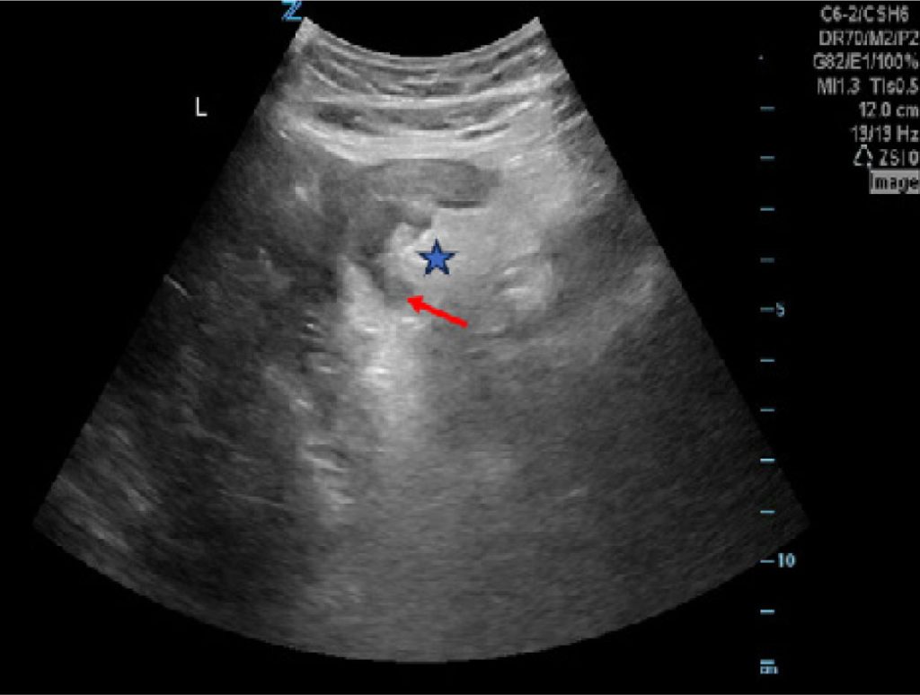
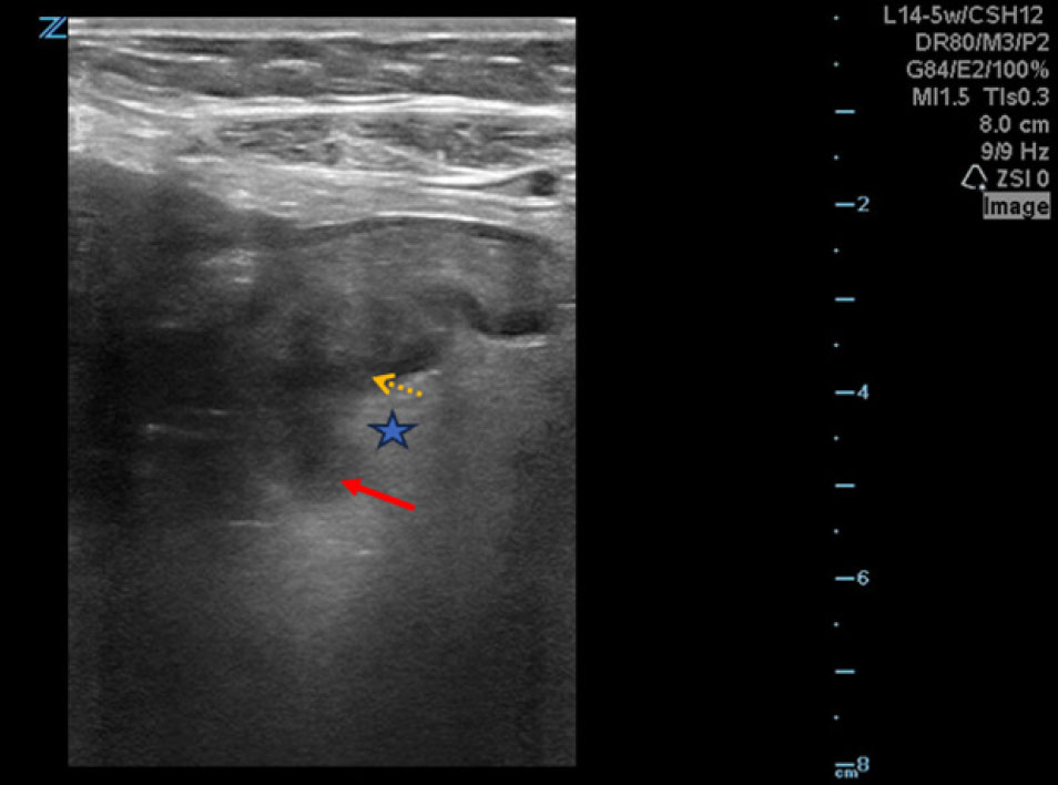
Given the high pretest clinical suspicion for diverticulitis, a bedside point-of-care ultrasound (POCUS) was performed using the curvilinear probe (Figure 1) followed by the linear probe to enhance the resolution (Figure 2), which showed hyperechoic pericolonic fat surrounding diverticula, areas of thickened colonic walls and sonographic tenderness of the area consistent with diverticulitis. After further discussion with the radiology attending, the CT interpretation was revised and confirmed our findings suggestive of proximal sigmoid diverticulitis. The patient was admitted to the hospital and was initiated on intravenous antibiotics. Six weeks later, she underwent a sigmoid colectomy for recurrent diverticulitis.

Figure 1. Ultrasound image obtained using a curvilinear probe demonstrates hyperechoic pericolonic fat (star) surrounding a diverticulum (solid red arrow)

Figure 2. Ultrasound image obtained using a linear probe showing hyperechoic pericolonic fat (star) surrounding a diverticulum (red arrow) with colonic wall thickening (dotted yellow arrow) with higher resolution

Discussion

In the setting of undifferentiated abdominal pain, CT is often relied upon as the diagnostic gold standard for patients in the emergency department, as it can differentiate many intra-abdominal pathologies and has a sensitivity of 97% and specificity of 98% for the diagnosis of acute colonic diverticulitis.1 In this scenario, we encountered a patient with a high pretest probability of diverticulitis. The initial CT interpretation failed to identify the pathology and it was only due to high clinical suspicion and our POCUS findings that the pathology was recognized. A discussion with radiology occurred, who recognized the pathology, changed the CT report and altered the patient’s treatment and disposition.

CT is widely recognized as the gold standard for the diagnosis of acute colonic diverticulitis. However, a meta-analysis of 630 patients did not show a statistical difference in accuracy between CT and ultrasound.2 Although POCUS is user-dependent, a more recent study demonstrated impressive accuracy of POCUS for diagnosing diverticulitis. As per Cohen et al., POCUS had a sensitivity of 92% and a specificity of 97%.3 Specifically, these metrics were found when all three of the following were seen: 1) bowel wall edema measuring 5 mm or greater surrounding an adjacent diverticulum, 2) enhancement of the surrounding pericolonic fat, and 3) sonographic tenderness to palpation.3 There is some diagnostic variability among sonographers for diverticulitis; some clinicians use a criterion of bowel thickness of at least 4mm.4 For Cohen et al., three of 14 false negative ultrasound scans had colonic wall thickness measurements less than 5mm of bowel wall thickness, but greater than 4mm.3 A 4mm cutoff would have improved POCUS sensitivity, although at the expense of specificity, as seen in a recent study by Shokoohi et al. that used a 4mm cutoff for bowel wall thickness resulting in a sensitivity of 95% and a specificity of 77%.4 Applying a wall thickness cutoff of 4mm may be more beneficial to increase sensitivity, identify more cases as a screening exam and confirm with CT.

Although more research is needed, POCUS has many benefits in the diagnosis of diverticulitis relative to CT. POCUS is a dynamic and interactive study in which the patient can help guide the sonographer for accurate visualization of the disease process. The performing clinician is also intimately aware of the patient’s clinical presentation, which can further direct the exam. It is feasible that POCUS could be applied in certain scenarios to avoid CT imaging and reduce radiation exposure. Despite being the gold standard, CT does not have 100% sensitivity, and it may be beneficial to consider ultrasound for the diagnosis of diverticulitis when clinical suspicion is high with a negative CT result.

Indications

  • Abdominal pain
  • Constipation
  • Diarrhea
  • Fever
  • Nausea and/or vomiting.

Technique

  • Position the patient supine. Consider having the patient bend their legs to soften the abdominal wall and allow for increased probe pressure.
  • Obtain images with either the linear, phased array or curvilinear probes. Given the wide footprint and improved penetration, a phased array or curvilinear probe is often used for the average adult patient.
  • A linear probe may allow sufficient depth penetration while having the advantage of a higher resolution for patients with lower body mass index (BMI).
  • Locate the area of interest by placing the probe on the area of maximal tenderness. Graded compression can be utilized to disperse obscuring bowel gas.
  • If no findings are seen in the area of interest, an expanded “lawn mower” approach can be utilized in which the probe is systematically moved up and down the abdomen with graded compression to evaluate all areas of the abdomen.

Pitfalls and Limitations

  • Images may be limited by body habitus or bowel gas, which may account for decreased sensitivity when compared to CT imaging.
  • Pneumoperitoneum may obscure evaluation of intraperitoneal structures.
  • Small bowel pathology can mimic large bowel diverticulitis, however it can be differentiated from the large bowel by the presence of plicae circulares or a series of folds that project into the small bowel lumen.
  • The presence of diverticulitis without visualized abscess or perforation does not rule out complicated diverticulitis.

References

  • Kandagatla, Pridvi G., and Amalia J. Stefanou. «Current status of the radiologic assessment of diverticular disease.» Clinics in colon and rectal surgery 31.04 (2018): 217-220
  • Laméris, Wytze, et al. «Graded compression ultrasonography and computed tomography in acute colonic diverticulitis: meta-analysis of test accuracy.» European radiology 18 (2008): 2498-2511.
  • Cohen, Allison, et al. «A prospective evaluation of point-of-care ultrasonographic diagnosis of diverticulitis in the emergency department.» Annals of Emergency Medicine 76.6 (2020): 757-766.
  • Shokoohi, Hamid, et al. «Accuracy of “TICS” ultrasound protocol in detecting simple and complicated diverticulitis: A prospective cohort study.» Academic Emergency Medicine 30.3 (2023): 172-179.