New York American College of Emergency Physicians

Penelope C. Lema, MD RDMS FACEP

Penelope C. Lema, MD RDMS FACEP

Vice Chair, Faculty Affairs Associate Professor, Department of Emergency Medicine Columbia University Vagelos College of Physicians & Surgeons

Guest Author Cullan Donnelly, MD

Guest Author Cullan Donnelly, MD

PGY- 2 Emergency Medicine Resident University of Rochester Medical Center Department of Emergency Medicine

John DeAngelis, MD FACEP Assistant Professor

John DeAngelis, MD FACEP Assistant Professor

Department of Emergency Medicine University of Rochester Medical Center

FOOSH, There It Is!

Case Presentation

A 17-year-old male with no significant past medical history presented to the emergency department after being a restrained driver in a motor vehicle accident. Airbags deployed, the patient was restrained, denied loss of consciousness and had full recollection of the events. The patient reported wrist pain and believes they jammed their outstretched wrists into the steering wheel or dashboard.

Physical examination revealed a superficial friction burn over the right anatomical snuffbox with tenderness to palpation in the same area and pain with axial loading of the right thumb. No snuffbox tenderness was noted in the left wrist. The patient’s vital signs were stable.

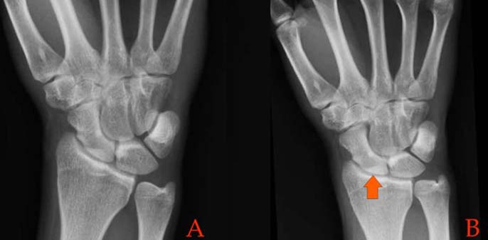
A X-ray of the right wrist showed no acute fracture or dislocation, growth plates are fused (Figure 1A).

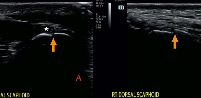
Point-of-care ultrasound (POCUS) of the wrist was performed with a L14-6s linear transducer (Mindray Te7, Mahwah, NJ, Shenzhen, China) that demonstrated cortical discontinuity along the dorsal and volar aspects of the scaphoid bone, concerning for radiographically silent scaphoid fracture, consistent with exam (Figures 2A and 2B).

The patient had minor burn care and was splinted in a thumb spica in a neutral position and was given an orthopedic referral and followed up seven days later with repeat X-rays. Repeat assessment in the orthopedic office one week later demonstrated continued snuffbox tenderness. The repeat X-ray (Figure 1B) revealed a fracture in the proximal third of the scaphoid that was identified by POCUS during the initial Emergency Department (ED) visit one week prior.

Discussions

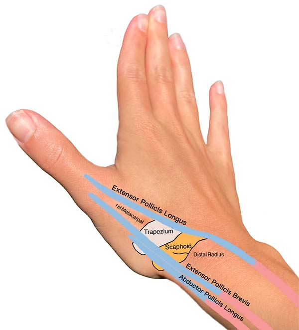
Scaphoid fractures have a propensity for malunion, non-union, chronic pain, arthritis, or even avascular necrosis due to the retrograde perfusion that the proximal end of the scaphoid receives.1 The most sensitive and widely used physical exam finding is called “anatomical snuffbox tenderness,” an anatomic space created by the adductor pollicis longus, extensor pollicis brevis and extensor pollicis longus on the radial aspect of the distal forearm which is where the dorsal aspect of the scaphoid bone can be palpated (Figure 3). When tenderness is encountered on physical exam, a fracture is assumed and a splint is placed with close outpatient follow-up to ensure the prevention of the morbidities associated with these fractures. Despite this common practice, it has been demonstrated in at least one retrospective study that up to 80% of splints applied were unnecessary due to a fracture not being found in subsequent imaging.2 Splints are not a benign application; they can limit a patient’s ability to work and perform activities of daily life. Plain film X-rays have only 60% sensitivity,3 and most departments do not regularly obtain CT or MRI imaging for a more definitive evaluation due to cost, time, or limited resources. The most sensitive but non-specific physical exam finding is anatomical snuffbox tenderness, but while the sensitivity is around 90%, the specificity is only around 40%.4 This is likely even worse if there is overlying skin injury such as an abrasion or burn, as in our case. Is there a cheaper, widely available, sensitive and specific imaging modality to identify scaphoid fractures? Scaphoid bone ultrasound has been shown to have a near 100% sensitivity and specificity when cortical disruption and effusion or hematoma are identified.5 In this case, the patient likely had trauma to the right wrist due to axial loading against the dashboard or steering wheel of the car during impact. As previously mentioned, a unique aspect of this situation is the fact he also had a burn over his wrist from the airbag, specifically over the anatomical snuffbox, making tenderness to palpation over this area significantly less specific than baseline. Pain with palpation over a superficial burn is to be expected even without an underlying injury beneath it. With the identification of cortical disruption of the scaphoid bone using ultrasound, both in the dorsal and volar views, further evidence was obtained to support a fracture requiring splinting and close orthopedic follow-up despite a negative wrist X-ray.

Indications

  • Fall onto outstretched hand
  • Deformity or swelling
  • Pain over anatomical snuffbox
  • Pain with axial loading of the thumb
  • Wrist Pain

Technique

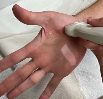
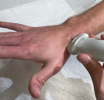
  • Use a linear ultrasound transducer on the dorsal and volar hand to obtain a sagittal view of the scaphoid (Figures 4 & 5). Transverse and oblique views should also be acquired to identify a fracture.
  • Use a generous amount of ultrasound gel and have the patient’s thumb in abduction and the wrist in ulnar deviation (as allowed by the patient’s pain and clinical scenario).
  • If the sonographic windows are too small, or if placement of the ultrasound transducer on the patient is limited due to body habitus or pain, consider using a water bath.
  • Once a sagittal view of the scaphoid bone is achieved, fan the transducer from side-to -side to scan through the bone to look for cortical disruption and/or hematoma.

Figure 1.(A) Initial xray and (B) repeat xray at one week demonstrating a subtle lucency in the proximal scaphoid consistent with a fracture (red arrow).

Figure 2.POCUS of the right wrist using a linear ultrasound transducer (A) ventral and (B) dorsal views of the scaphoid of the right wrist. A fracture is identified with cortical discontinuity (orange arrows) and lipohemarthrosis (white star)

Pitfalls and Limitations

  • Patient’s level of pain, cooperation and body habitus.
  • False positives due to osteophytes, vascular channels mimicking disruptions in bony cortex, or periosteal hematoma/swelling without cortical disruption.
  • This technique with POCUS has not yet been adopted as the “gold standard” to rule out the requirement for splinting and orthopedic follow-up. If the patient has anatomical snuffbox tenderness, recommend treatment as per the current standard of care. However, POCUS can provide additional diagnostic information that an occult fracture is present not initially identified on X-ray.

Pearls

  • The physical exam is key for fall onto an outstretched hand (FOOSH ) injuries.
  • There is a high rate of missed scaphoid fractures, however, up to 80% of splints are unnecessary in retroactive studies using follow-up imaging.
  • Ultrasound of the scaphoid bone with cortical disruption and effusion present has a 100% sensitivity and specificity.
  • This is not likely to change overall practice or decrease the number of splints applied in the short term, however, it can provide reassurance we are splinting a fracture. With additional research, it may be promising, with sufficient high sensitivity, to rule out fractures in the future.

References

  • Schmid GL, Lippmann S, Unverzagt S, Hofmann C, Deutsch T, Frese T. The Investigation of Suspected Fracture-a Comparison of Ultrasound With Conventional Imaging. Dtsch Arztebl Int. 2017 Nov 10;114(45):757-764. doi: 10.3238/ arztebl.2017.0757. PMID: 29202925; PMCID: PMC5729224.
  • Nguyen Q, Chaudhry S, Sloan R, Bhoora I, Willard C. The clinical scaphoid fracture: early computed tomography as a practical approach. Ann R Coll Surg Engl. 2008 Sep;90(6):488-91. doi: 10.1308/003588408X300948. Epub 2008 Jul 2. PMID: 18598597; PMCID: PMC2647242.
  • Tiel-van Buul MM, van Beek EJ, Borm JJ, Gubler FM, Broekhuizen AH, van Royen EA. The value of radiographs and bone scintigraphy in suspected scaphoid fracture. A statistical analysis. J Hand Surg Br 1993; 18: 403–6.
  • Freeland P. Scaphoid tubercle tenderness: a better indicator of scaphoid fractures? Arch Emerg Med. 1989 Mar;6(1):46-50. doi: 10.1136/emj.6.1.46. PMID:2712988; PMCID: PMC1285557.
  • Ortiz, A. What is the role of ultrasonography in the early diagnosis of scaphoid fractures?, European Journal of Radiology Open, Volume 8, 2021, 100358, ISSN 2352-0477, https://doi.org/10.1016/j.ejro.2021.100358.

Figure 3. General anatomy of the “anatomical snuffbox,” with the location of the scaphoid bone.

Figure 4. Placement of the linear ultrasound transducer over the scaphoid at the volar aspect of the wrist.

Figure 5. Placement of the linear ultrasound transducer over the scaphoid at the dorsal aspect of the wrist.