New York American College of Emergency Physicians

Penelope C. Lema, MD RDMS FACEP

Penelope C. Lema, MD RDMS FACEP

Vice Chair, Faculty Affairs Associate Professor, Department of Emergency Medicine Columbia University Vagelos College of Physicians & Surgeons

Guest Author Di Coneybeare, MD MHPE FPD-AEMUS

Guest Author Di Coneybeare, MD MHPE FPD-AEMUS

Director, Emergency Ultrasound Fellowship Assistant Professor of Emergency Medicine Columbia University Vagelos College of Physicians and Surgeons Department of Emergency Medicine

Guest Author Bannet Muhoozi, MD

Guest Author Bannet Muhoozi, MD

Emergency Ultrasound Fellow Clinical Instructor of Emergency Medicine Columbia University Vagelos College of Physicians and Surgeons Department of Emergency Medicine

Significance of Mitral Valve Prolapse on Cardiac Point of Care Ultrasound (POCUS) in the Emergency Department

Case

A 77-year-old female with a medical history of hypertension and hyperlipidemia presented to the Emergency Department (ED) after a syncopal episode with pressure-like chest pain radiating to the left arm. She had similar chest pain intermittently for months, but began having palpitations, shortness of breath and lightheadedness the morning of presentation. While walking her dog, she had a syncopal episode and EMS was called. On arrival to the ED, vital signs were normal without hypotension or tachycardia. The EKG demonstrated a LBBB and ST depression in lead II, similar to a prior one from two years ago.

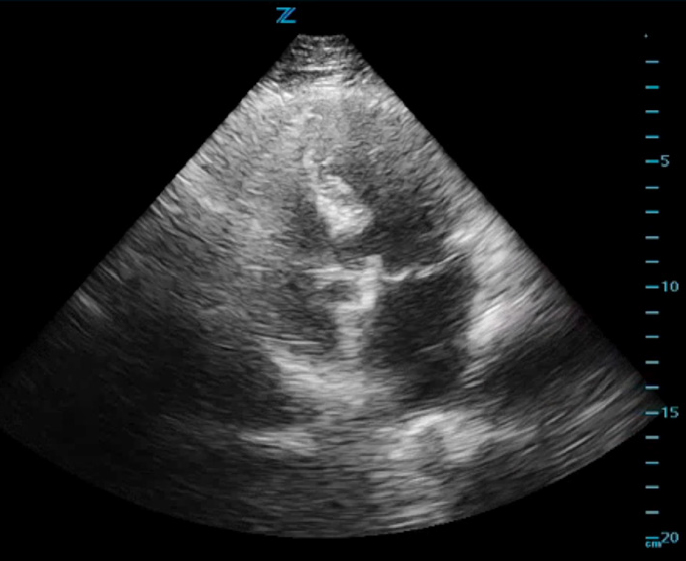
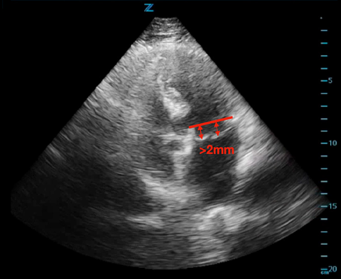
ED cardiac point-of-care ultrasound (POCUS) revealed mild left ventricular sigmoid hypertrophy and mitral valve prolapse of both anterior and posterior valves with mitral regurgitation (Figures 1A and 1B). There was normal systolic left ventricular function without segmental wall motion abnormalities or pericardial effusion. There were no anterior or lateral lung B-lines demonstrating pulmonary vascular congestion. The serum troponin level was mildly elevated and remained stable on repeat evaluation.

Consultation with cardiology was initiated due to the ED POCUS findings of mitral valve prolapse and increased suspicion for cardiac dysrhythmia, such as rapid atrial fibrillation, contributing to the patient’s syncope. In conjunction with cardiology recommendations, the patient was initiated on anticoagulation due to suspicion for atrial fibrillation and was admitted to the cardiology service. The patient subsequently underwent cardiac catheterization which showed non-obstructive coronary disease. Inpatient echocardiogram showed a myxomatous mitral valve with moderate prolapse of both leaflets and moderate mitral regurgitation consistent with ED POCUS. She was started on an optimized cardiac medication regimen, including aspirin, atorvastatin and metoprolol. She remained chest pain free and hemodynamically stable during the hospital course and was discharged home with cardiology follow up the next day. This case highlights the crucial role of POCUS in helping to identify the underlying cause of syncope and diagnosing symptomatic mitral valve prolapse.

Discussions

Mitral valve prolapse (MVP) is classified as primary, from spontaneous tissue disease such as myxomatous degeneration or secondary, when due to an underlying disorder like Marfans or other connective tissue disease. Furthermore, MVP causes primary mitral regurgitation (MR), due to mitral valve leaflet pathology, as opposed to secondary mitral regurgitation from left ventricular dysfunction.1,2

MVP often presents with symptoms that can mimic more common cardiac pathologies, like acute coronary syndrome (ACS). Although historically associated with a range of symptoms—such as chest pain and palpitations—most MVP patients are asymptomatic.3,4 When symptoms do occur, they usually stem from significant MR or cardiac arrhythmias.3,4 The condition is often identified either through echocardiography prompted by unrelated medical evaluations or auscultatory findings, rather than symptoms alone. Given its varying presentation it is an important differential diagnosis in patients who present to the ED with respiratory distress, chest pain and syncope.

Clinically, MVP is diagnosed via specific echocardiographic criteria, including thickened and redundant mitral leaflets that billow in systole at least 2 mm below the plane of the mitral annulus, towards the atrium. MR may or may not accompany these findings.5 It’s important to clarify that myocardial ischemia does not cause MVP. However, during a myocardial infarction, a ruptured papillary muscle may lead to mitral valve leaflets sagging into the left atrium causing mitral regurgitation.

In our case the patient was at high risk for ACS given her age, history and symptomatology. However, cardiac POCUS revealed another underlying issue providing an alternative differential diagnosis to ACS that could explain her syncopal episode, dyspnea and chest pain. This underscores the utility of POCUS to evaluate patients with non-specific cardiac symptoms, such as chest pain or shortness of breath, though it doesn’t eliminate the need for a coronary catheterization.

In conclusion, our case underscores the potential of cardiac POCUS as a versatile diagnostic tool in the emergency setting. This case emphasizes the need for emergency physicians to be proficient in the use of POCUS not only for ruling out life-threatening conditions but also for recognizing other structural abnormalities like MVP that could significantly influence clinical decision-making.

  • Cardiac Murmur
  • Chest pain
  • Palpitations
  • Shortness of breath
  • Tachycardia

Technique

  • Place the patient in a supine position. Consider having them lay on their left side to bring the heart closer to the chest wall.
  • Use a phased array probe that allows for adequate penetration between the ribs. The mitral valve is best captured in either the apical four chamber or parasternal long axis view. Please refer to
    New York ACEP EPIC volume 32:02:14, pages 6 – 8.
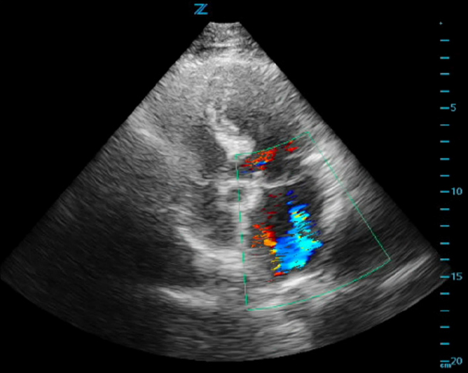
  • Use color flow Doppler to assess for the presence and severity of mitral regurgitation. Variables such as the angle of insonation, depth and sample size are crucial for an accurate assessment. MVP is defined as systolic billowing of one or both mitral leaflets ≥2 mm below the mitral annular plane into the left atrium. This is often seen in a long-axis view, either parasternal or apical four-chamber (figure 2).
  • Moderate mitral regurgitation features a broad and sizable jet that takes up a portion of the left atrium. In severe MR, the doppler jets reach the base of the left atrium or fill more than 40% of its area.
  • M-mode echocardiography is outdated and should not be used for diagnosing MVP. Normal motion at the base may mask or mimic MVP

References

  • Otto CM, Nishimura RA, Bonow RO, Carabello BA, Erwin JP 3rd, Gentile F, Jneid H, Krieger EV, Mack M, McLeod C, O’Gara PT, Rigolin VH, Sundt TM 3rd, Thompson A, Toly C. 2020 ACC/AHA Guideline for the Management of Patients With Valvular Heart Disease: A Report of the American College of Cardiology/American Heart Association Joint Committee on Clinical Practice Guidelines. Circulation. 2021 Feb 2;143(5):e72-e227. doi: 10.1161/CIR.0000000000000923. Epub 2020 Dec 17. Erratum in: Circulation. 2021 Feb 2;143(5):e229. Erratum in: Circulation. 2023 Aug 22;148(8):e8. PMID: 33332150.
  • Vahanian A, Beyersdorf F, Praz F, Milojevic M, Baldus S, Bauersachs J, Capodanno D, Conradi L, De Bonis M, De Paulis R, Delgado V, Freemantle N, Gilard M, Haugaa KH, Jeppsson A, Jüni P, Pierard L, Prendergast BD, Sádaba JR, Tribouilloy C, Wojakowski W; ESC/EACTS Scientific Document Group. 2021 ESC/EACTS Guidelines for the management of valvular heart disease. Eur Heart J. 2022 Feb 12;43(7):561-632. doi: 10.1093/eurheartj/ehab395. Erratum in: Eur Heart J. 2022 Feb 18;: PMID: 34453165.
  • Devereux RB, Kramer-Fox R, Brown WT, Shear MK, Hartman N, Kligfield P, Lutas EM, Spitzer MC, Litwin SD. Relation between clinical features of the mitral prolapse syndrome and echocardiographically documented mitral valve prolapse. J Am Coll Cardiol. 1986 Oct;8(4):763- 72. doi: 10.1016/s0735-1097(86)80415-6. PMID: 3760352.
  • Freed LA, Levy D, Levine RA, Larson MG, Evans JC, Fuller DL, Lehman B, Benjamin EJ. Prevalence and clinical outcome of mitral-valve prolapse. N Engl J Med. 1999 Jul 1;341(1):1-7. doi: 10.1056/NEJM199907013410101. PMID: 10387935.
  • Bonow RO, Carabello BA, Chatterjee K, de Leon AC Jr, Faxon DP, Freed MD, Gaasch WH, Lytle BW, Nishimura RA, O’Gara PT, O’Rourke RA, Otto CM, Shah PM, Shanewise JS; 2006 Writing Committee Members; American College of Cardiology/American Heart Association Task Force. 2008 Focused update incorporated into the ACC/AHA 2006 guidelines for the management of patients with valvular heart disease: a report of the American College of Cardiology/American Heart Association Task Force on Practice Guidelines (Writing Committee to Revise the 1998 Guidelines for the Management of Patients With Valvular Heart Disease): endorsed by the Society of Cardiovascular Anesthesiologists, Society for Cardiovascular Angiography and Interventions, and Society of Thoracic Surgeons. Circulation. 2008 Oct 7;118(15):e523-661. doi: 10.1161/CIRCULATIONAHA.108.190748. Epub 2008 Sep 26. PMID: 18820172.
  • Grewal J, Suri R, Mankad S, Tanaka A, Mahoney DW, Schaff HV, Miller FA, Enriquez-Sarano M. Mitral annular dynamics in myxomatous valve disease: new insights with real-time 3-dimensional echocardiography. Circulation. 2010 Mar 30;121(12):1423-31. doi: 10.1161/CIRCULATIONAHA.109.901181. Epub 2010 Mar 15. PMID: 20231533.
  • Morningstar JE, Nieman A, Wang C, Beck T, Harvey A, Norris RA. Mitral Valve Prolapse and Its Motley Crew-Syndromic Prevalence, Pathophysiology, and Progression of a Common Heart Condition. J Am Heart Assoc. 2021 Jul 6;10(13):e020919. doi: 10.1161/JAHA.121.020919. Epub 2021 Jun 22. PMID: 34155898; PMCID: PMC8403286.

Figures 1A. B-mode ultrasound image of the apical four chamber view with mitral valve prolapse

Figure 1B. Mitral valve prolapse (red arrows) with mitral leaflets ≥2 mm below the mitral annular plane into the left atrium.

Figure 2. Ultrasound image of the cardiac apical four chamber view demonstrating mitral valve regurgitation with color flow Doppler.